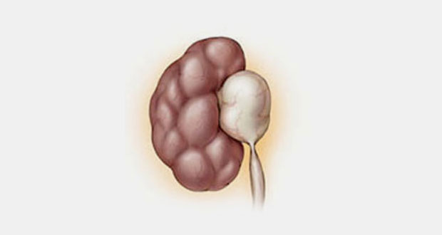
Pelvi-ureteric junction obstruction (PUJ obstruction)
Idiopathic PUJ obstruction
- More common in men
- Affects left kidney more often than right
- 10% cases are bilateral
- Cause is unknown but important factors may be: abnormal lower pole vessels; persistant foetal urothelial fold
- Usually presents in adolescence or early adult life
- Presenting symptom may be loin pain – worse after alcohol
- In late cases a renal mass may be palpable
- Haematuria is an uncommon feature
- 10% develop UTIs (urinary tract infection) and 3% renal colic (pain)
- Diagnosis can be confirmed by ultrasound
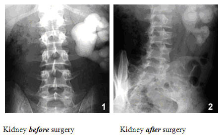
- IVU or retrograde ureterogram show a classical appearance
- Isotope renography allows assessment of percentage of renal function
Surgical Intervention for PUJ Obstruction
If the transition from the renal pelvis to the ureter is narrow, the urine will not drain easily and backs up causing dilatation of the collecting system proximal to that point and enlargement of the renal pelvis. This dilatation of the collecting system is referred to as hydronephrosis.
The aims of treatment are to:
- Relieve symptoms
- Preserve renal function
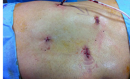
Now pyeloplasty is done laparoscopically by Dr.Ramesh at Apollo Hospitals, Chennai as minimally invasive procedure.
Current advancement in treatment of PUJ obstruction is Robotic assited Laparoscopic Pyeloplasty.
- Precise mucosa to mucosa anastomosis
- Useful in redo surgery
Currently minimally invasive treatment like endopyelotomy is also available.
 Urologist in Chennai | Robotic Urologist in India | Chennai Urology


